Vitamin D Pg Ml
Reference Range
The biologically active form of vitamin D is 1,25-dihydroxyvitamin D (1,25(OH)2 D). Measurement of serum levels of 1,25(OH)2 D should be considered upon suspicion of deficiency or excess of this form of the vitamin.
Reference ranges for 1,25(OH)2 D may be reported as either pg/mL or pmol/L. The molecular weight of 1,25(OH)2 D is approximately 416.7, yielding the following conversion factors: 1 pmol/L = 0.42 pg/mL; conversely, 1 pg/mL = 2.4 pmol/L.
Reference ranges for 1,25(OH)2 D are as follows [1] :
-
Males: 18-64 pg/mL
-
Females: 18-78 pg/mL
![]()
Interpretation
Plasma 1,25-dihydroxyvitamin D (1,25(OH)2 D) is tightly controlled by plasma parathyroid hormone (PTH), serum calcium, serum phosphate, and fibroblast-like growth factor 23 (FGF-23).
Decreased 1,25-dihydroxyvitamin D levels
Decreased levels of 1,25(OH)2 D can result from chronic kidney disease, various heritable disorders, tumor-induced osteomalacia, the use of HIV protease inhibitors, or severe vitamin D deficiency.
Chronic kidney disease: Low 1,25(OH)2 D levels have been shown to present even in early stages of kidney failure. The decrease of 1,25(OH)2 D level is more prominent when kidney failure progresses. In a study by Levin et al (2007), 13% of patients with an estimated glomerular filtration rate (eGFR) greater than 80 mL/min and more than 60% of patients with an eGFR of less than 30 mL/min had low serum levels of 1,25(OH)2 D. [2] Impaired production of the enzyme 1α-hydroxylase in kidney failure was thought to be the main mechanism. However, phosphate retention and FGF-23 also contribute to the decreased synthesis of 1,25(OH)2 D. [3]
Heritable disorders associated with low 1,25(OH)2 D levels include vitamin D–dependent rickets type 1 (inactivating mutation in the 1-hydroxylase gene), [4] autosomal-dominant hypophosphatemic rickets (mutation of the gene coding for FGF-23, which prevents its breakdown), [5] and X-linked hypophosphatemic rickets (mutations that elevate levels of FGF-23). [6]
In tumor-induced osteomalacia, tumor-secreted FGF-23 inhibits enzyme 1α-hydroxylase and subsequently results in decreased 1,25(OH)2 D synthesis. [7]
HIV protease inhibitors have been reported to markedly suppress the activities of 25- and 1α-hydroxylase and thus affect 1,25(OH)2 D synthesis. [8] In a cohort study including 671 patients, progression to bone demineralization was observed in 28% of the patients over a median of 2.5 years. Patients who were concurrently using protease inhibitors were at greater risk for worsening bone demineralization than those who were not using protease inhibitors (OR 1.64; 95% CI, 1.35-2.04; P< 0.0001). [9]
Severe vitamin D deficiency: 25(OH)D is the main substrate of 1,25(OH)2 D. Vitamin D deficiency can affect the production of 1,25(OH)2 D owing to the lack of substrate. A positive correlation between serum levels of 25(OH)D and 1,25(OH)2 D was observed during seasonal changes. Treatment with 25(OH)D can normalize 1,25(OH)2 D concentrations in patients with vitamin D deficiency. [10]
Increased 1,25-dihydroxyvitamin D levels
Increased 1,25(OH)2 D levels can result from extrarenal 1α-hydroxylation or hereditary vitamin D–resistant rickets.
In granulomatous disease such as lymphoproliferative disorders, sarcoidosis, tuberculosis, and inflammatory bowel disease, 1α-hydroxylase enzyme activity was found in macrophages as the extrarenal source of 1,25(OH)2 D. When 1α-hydroxylase is activated, it converts 25(OH)D to 1,25(OH)2 D, just as what occurs under physiologic conditions in the kidneys. [11] However, unlike the kidney, the 1α-hydroxylase in the macrophages in granulomatous diseases is not controlled by the usual physiologic regulators. Moreover, not all conditions or all patients with increased macrophage activity manifest increases in 1α-hydroxylase activity. In vitro studies of monocytes/macrophages indicate that gamma interferon is an important regulator of 1α-hydroxylase but only when other key signaling pathways are also activated (eg, JAK-STAT and MAP-Kinase). [12]
Hereditary vitamin D-resistant rickets is a very rare autosomal recessive disorder in which mutations of vitamin D receptor (VDR) coding genes cause failure or abnormal binding of vitamin D to VDRs. [13, 14] Patients usually present with hypocalcemia, early-onset rickets, alopecia, and other ectodermal anomalies.
![]()
Collection and Panels
Specimen: Blood (0.25 mL room-temperature serum)
Container: Red-top tube, serum separator tube (also acceptable: lavender [EDTA] or pink [K2 EDTA])
Collection method: Routine venipuncture
Storage/transport temperature: Refrigerated (Note: Some laboratories require a frozen specimen; one should check with the specific laboratory before ordering the test)
Stability (collection to initiation of testing; after separation from cells): Ambient, 72 hours; refrigerated, 1 week; frozen, 6 months
Panels: 1,25(OH)2 D is not included in routine comprehensive metabolic panels; it should be ordered as a separate blood test
Other instructions: Collect blood in a standard red-top tube; allow blood to clot at room temperature; centrifuge and separate the serum from the cells immediately within 2 hours of collection
Assays for serum 1,25(OH)2D
Compared to 25(OH)D, 1,25(OH)2 D circulates in the human body at a very low concentration, making its serum levels challenging to assess.
Radioreceptor assay
First introduced in 1974, the radioreceptor assay (RRA) originally included several purification steps. Chicken intestinal VDR was used as the binding agent. [15] In 1984, Reinhardt et al presented a significant improvement in the RRA method whereby the high performance liquid chromatography step was eliminated. Measurement of 1,25(OH)2 D was achieved by a nonequilibrium assay using 1,25(OH)2 D receptor from calf thymus. [16]
This modification of RRA has made it a more practical assay. However, it still has some disadvantages, including the necessity of VDR isolation and the limitations that comes from using3 H-1,25(OH)2 D3 as the reporter. [17]
Radioimmunoassay
The first radioimmunoassay (RIA) that measured 1,25(OH)2 D was introduced in 1978. [18] Over the years, an RIA for 1,25(OH)2 D using a radio-iodinated (125 I) tracer was developed by Hollis et al. [19] The assay involves acetonitrile extraction, treatment of the crude extract supernatant with sodium periodate, extraction and purification of endogenous 1,25(OH)2 D by solid-phase chromatography, and quantification by RIA.
![]()
Background
Description
Vitamin D is a group of fat-soluble compounds with a four-ringed cholesterol backbone; it is now recognized as a prohormone.
Vitamin D exists in two major forms: vitamin D2 (ergocalciferol) and vitamin D3 (cholecalciferol). The precursor for vitamin D2 is a plant sterol ergosterol. D2 can be synthesized by ultraviolet irradiation of ergosterol from yeast. Similarly, vitamin D3 is synthesized in the body when sunlight (ultraviolet B, wavelength 280-315 nm) photoisomerizes 7-dehydroxycholesterol found in the skin. D3 is also found in animal-based foods (eg, fatty fish, liver, milk, eggs).
Vitamin D2 and D3, regardless of the source, are biologically inactive. They are transformed into the biologically active molecule 1,25 dihydroxyvitamin D. After being synthesized in the skin or absorbed (in chylomicrons) from the gastrointestinal (GI) tract, most vitamin D is bound to specific carrier proteins in the blood (vitamin D–binding protein [DBP] and albumin) and transported to the liver. In the liver, vitamin D is hydroxylated by the enzyme 25-hydroxylase (CYP2R1) to become 25(OH)D. 25(OH)D is the major circulating form of vitamin D. From the liver, 25(OH)D is transported to the kidneys via the same carrier proteins as above.
1,25 dihydroxyvitamin D (1,25(OH)2 D) is formed when 25(OH)D is hydroxylated by the enzyme 1α-hydroxylase (CYP27B1), which is located in the mitochondria of proximal tubules of the kidney. 1,25(OH)2 D is the biologically active form of vitamin D. As a result of 1 and 25 hydroxylation, the prohormone vitamin D has been transformed into an active hormone.
1,25(OH)2 D is a steroidlike hormone. In target cells, such as classic steroid hormones, it binds to a specific cytoplasmic VDR; the vitamin D bound to VDR then translocates to the nucleus, where its effects are initiated at a transcriptional level. [20] The main established actions of 1,25(OH)2 D collectively increase calcium in the body and modulate the skeleton. It increases the intestinal absorption of calcium and phosphate, decreases renal excretion of calcium and phosphate, suppresses PTH production, and regulates osteoblast function and bone resorption. It has been suggested that 1,25(OH)2 D has roles beyond the calcium-skeletal axis. [20]
The identification of VDRs in various cells has prompted the investigation of vitamin D in immunomodulation, cancer prevention and therapy, autoimmune disease, cardiovascular disease, and other nonskeletal organs.
The synthesis of 1,25(OH)2 D is tightly regulated by PTH, serum calcium, serum phosphate, and fibroblast-like growth factor 23 (FGF-23). Increased levels of PTH and hypophosphatemia stimulate the enzyme 1α-hydroxylase and, subsequently, the synthesis of 1,25(OH)2 D. FGF-23 is a circulating hormone synthesized by osteocytes and osteoblasts. 1,25(OH)2 D and phosphate intake stimulates the synthesis of FGF-23, which, in turn, inhibits 1,25(OH)2 D production, reduces the expression of renal sodium–phosphate transporters, and activates the metabolizing of active 1,25(OH)2 D to the inactive metabolite 24,25(OH)2 D. [21]
As a fat-soluble molecule, vitamin D is stored in adipose tissue; however, the exact mechanism by which vitamin D is regulated and mobilized from adipose tissue has not been elucidated at this time. Most vitamin D products are excreted through bile into the gut. Very little is eliminated via kidneys (see graph below).
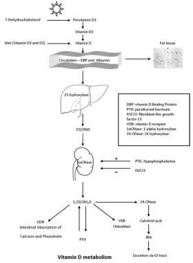
Vitamin D metabolism.
Indications/Applications
Measuring serum levels of 1,25(OH)2 D should be considered upon suspicion of deficiency or excess of 1,25(OH)2 D.
1,25(OH)2 D assessment may be beneficial in patients with chronic kidney failure; early-onset rickets; family history of rickets; or long-term use of protease inhibitors, glucocorticoids, or anticonvulsants who are at risk of hypocalcemia from low concentration of 1,25(OH)2 D.
1,25(OH)2 D may be helpful in diagnosing parathyroid function disorders. A high serum level of 1,25(OH)2 D, for example, may suggest of primary hyperparathyroidism, whereas a normal or low serum level is more likely found in secondary hyperparathyroidism.
Patients with hypercalcemia can undergo testing for both 25(OH)D and 1,25(OH)2 D to rule out vitamin D intoxication or disorders that can enhance 1,25(OH)2 D synthesis, such as lymphoma, sarcoidosis, and other granulomatous diseases.
Serial 1,25(OH)2 D levels can be used in monitoring the efficacy of treatment in patients receiving 1,25(OH)2 D supplementation.
In overweight and obese individuals, serum 1,25(OH)2 D concentrations were reported to be correlated with 25(OH)D levels and seasonal variations. This suggests that measurement of both levels might be beneficial when assessing vitamin D stores in this population. [22]
Considerations
The half-life of 1,25(OH)2 D is approximately 4 hours. Therefore, it is not useful in assessing the total-body vitamin D status. Also, it must be noted that a normal level of 1,25(OH)2 D does not exclude a diagnosis of vitamin D deficiency. Measurement of 25(OH)D levels should be used for this purpose.
![]()
-
Pagana KD, Pagana TJ, Pagana TN. Mosby's Diagnostic & Laboratory Test Reference. 14th ed. St. Louis, Mo: Elsevier; 2019.
-
Levin A, Bakris GL, Molitch M, Smulders M, Tian J, Williams LA, et al. Prevalence of abnormal serum vitamin D, PTH, calcium, and phosphorus in patients with chronic kidney disease: results of the study to evaluate early kidney disease. Kidney International. 2007. 71(1):31-8. [Medline]. [Full Text].
-
Gutierrez O, Isakova T, Rhee E, Shah A, Holmes J, Collerone G, et al. Fibroblast growth factor-23 mitigates hyperphosphatemia but accentuates calcitriol deficiency in chronic kidney disease. J Am Soc Nephrol. July 2005. 16(7):2205-15. [Medline]. [Full Text].
-
Kitanaka S, Takeyama K, Murayama A, Kato S. The molecular basis of vitamin D-dependent rickets type I. Endocr J. 2001. 48(4):427. [Medline].
-
White KE, Jonsson KB, Carn G, Hampson G, Spector TD, Mannstadt M, et al. The autosomal dominant hypophosphatemic rickets (ADHR) gene is a secreted polypeptide over expressed by tumors that cause phosphate wasting. J Clin Endocrinol Metab. 2001 Feb. 86(2):497-500. [Medline]. [Full Text].
-
Ruppe MD, Brosnan PG, Au KS, Tran PX, Dominguez BW, Northrup H. Mutational analysis of PHEX, FGF23 and DMP1 in a cohort of patients with hypophosphatemic rickets. Clin Endocrinol (Oxf). 2011 Mar. 74(3):312-8. [Medline]. [Full Text].
-
Habra MA, Jimenez C, Huang SC, Cote GJ, Murphy WA Jr, Gagel RF, et al. Expression analysis of fibroblast growth factor-23, matrix extracellular phosphoglycoprotein, secreted frizzled-related protein-4, and fibroblast growth factor-7: identification of fibroblast growth factor-23 and matrix extracellular phosphoglycoprotein as major factors involved in tumor-induced osteomalacia. Endocrine Practice. 2008 Dec. 14(9):1108-14. [Medline]. [Full Text].
-
Cozzolino M, Vidal M, Arcidiacono MV, Tebas P, Yarasheski KE, Dusso AS. Dusso AS HIV-protease inhibitors impair vitamin D bioactivation to 1,25-dihydroxyvitamin D. AIDS. 2003 Mar 7. 17(4):513-20. [Medline]. [Full Text].
-
Bonjoch A, Figueras M, Estany C, Perez-Alvarez N, Rosales J, del Rio L, et al. High prevalence of and progression to low bone mineral density in HIV-infected patients: a longitudinal cohort study. AIDS. 2010 Nov 27. 24(18):2827-33. [Medline].
-
Bouillon RA, Auwerx JH, Lissens WD, Pelemans WK. Vitamin D status in the elderly: seasonal substrate deficiency causes 1,25-dihydroxycholecalciferol deficiency. Am J Clin Nutr. 1987 Apr. 45(4):755-63. [Medline]. [Full Text].
-
Inui N, Murayama A, Sasaki S, Suda T, Chida K, Kato S, et al. Correlation between 25-hydroxyvitamin D3 1 alpha-hydroxylase gene expression in alveolar macrophages and the activity of sarcoidosis. Am J Med. 2001. 110(9):687-93. [Medline]. [Full Text].
-
Lut Overbergh, Katinka Stoffels,Mark Waer, Annemieke Verstuyf, Roger Bouillon, and Chantal Mathieu. Immune Regulation of 25-Hydroxyvitamin D-1- alpha Hydroxylase in Human Monocytic THP1 Cells: Mechanisms of Interferon-Mediated Induction. J Clin Endocrinol Metab. 2006 Sep. 91(9):3566-74. [Medline]. [Full Text].
-
Malloy PJ, Feldman D. Genetic disorders and defects in vitamin d action. Endocrinol Metab Clin North Am. 2010. 39(2):333-36. [Medline]. [Full Text].
-
Malloy PJ, Eccleshall TR, Gross C, Van Maldergem L, Bouillon R, Feldman D. D Hereditary vitamin D resistant rickets caused by a novel mutation in the vitamin D receptor that results in decreased affinity for hormone and cellular hyporesponsiveness. J Clin Invest. 1997 Jan. 99(2):297-304. [Medline]. [Full Text].
-
Brumbaugh PF, Haussler DH, Bressler R, Haussler MR. Radioreceptor assay for 1α,25-dihydroxyvitamin D3. Science ;183:. 1974. 183(4129):1089–1091. [Medline].
-
Reinhardt TA, Horst RL, Orf JW, Hollis BW. A microassay for 1,25-dihydroxyvitamin D notrequiring high performance liquid chromatography: Application to clinical studies. Journal of Clinical Endocrinology and Metabolism ;. 1984. 58(1):91–98. [Medline]. [Full Text].
-
Bruce W. Hollis and Ronald L. Horst. The Assessment of Circulating 25(OH)D and 1,25(OH)2D: Where We Are and Where We Are Going. J Steroid Biochem Mol Biol. 2007 March. 103(3-5):473–476. [Medline]. [Full Text].
-
Clemens TL, Hendy GN, Graham RF, Baggiolini EG, Uskokovic MR, O'Riordan JLH. A radioimmunoassay for 1,25-dihydroxycholecalciferol. Clinical Science and Molecular Medicine. 1978. 54(3):329–332. [Medline].
-
Hollis BW, Kamerud JQ, Kurkowski A, Beaulieu J, Napoli JL. Quantification of circulating 1,25- dihydroxyvitamin D by radioimmunoassay with 125I-labeled tracer. Clinical Chemistry. 1996;. 42(4):586–592. [Medline]. [Full Text].
-
B. Maestro, S. Molero, S. Bajo, N. D´avila, and C. Calle. Transcriptional activation of the human insulin receptorgene by 1,25-dihydroxyvitamin D3. Cell Biochemistry and Function. 2002. 20(3):227–232. [Medline]. [Full Text].
-
AU PriéD, Friedlander G SO. Reciprocal control of 1,25-dihydroxyvitamin D and FGF23 formation involving the FGF23/Klotho system. Clin J Am Soc Nephrol. 2010. 5(9):1717-22. [Medline]. [Full Text].
-
Lagunova Z, Porojnicu AC, Vieth R, Lindberg FA, Hexeberg S, Moan J. Serum 25-hydroxyvitamin D is a predictor of serum 1,25-dihydroxyvitamin D in overweight and obese patients. J Nutr. 2011 Jan. 141(1):112-7. [Medline].
-
Vitamin D metabolism.

Source: https://emedicine.medscape.com/article/2088672-overview#:~:text=Reference%20ranges%20for%201%2C25(OH)2%20D%20are,%3A%2018%2D78%20pg%2FmL
